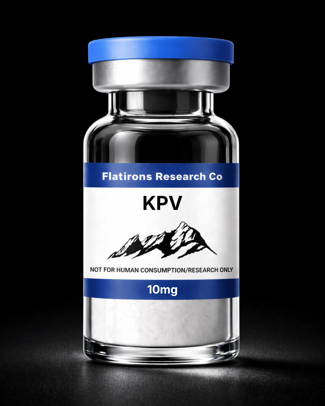
KPV, 10mg
$33.00
- Product Name: KPV
- Sequence: Lys-Pro-Val
- Molecular Formula: C16H30N4O4
- Molecular Weight: 342.44 g/mol
- Research Only: Yes
- Form: Lyophilized Solid
- Purity: 99%
- Storage: Keep refrigerated upon reconstitution
Some COAs may contain redactions of confidential or proprietary information. These redactions do not impact or obscure any analytical data, including compound identity, purity, or test results.
Availability: 16 in stock
Bulk Pricing
- Buy 1 - 2 vials for $33.00 each
- Buy 3 - 4 vials for $31.35 each (5% off)
- Buy 5 - 7 vials for $29.70 each (10% off)
- Buy 8 - 11 vials for $29.04 each (12% off)
- Buy 12 - 15 vials for $28.05 each (15% off)
- Buy 16+ vials for $26.40 each (20% off)
KPV (Lys-Pro-Val) is a C-terminal tripeptide fragment derived from alpha-melanocyte-stimulating hormone (α-MSH, residues 11-13). Published research has investigated KPV in a variety of experimental signaling and inflammatory pathway models, where it has demonstrated melanocortin receptor-independent activity distinct from its parent peptide.
Molecular Signaling & Transport Mechanisms
Experimental findings indicate that KPV is transported intracellularly through the PepT1 oligopeptide transporter, exhibiting measurable affinity in intestinal epithelial cell models. In vitro studies have reported intracellular accumulation and modulation of NFκB-associated signaling pathways through interference with p65RelA/importin-α3 interactions, resulting in altered nuclear translocation dynamics.
Additional published investigations have evaluated KPV’s influence on MAPK signaling cascades, including ERK1/2, JNK, and p38 pathways in epithelial cell systems. Observed effects occurred independently of classical melanocortin receptor activation and were associated with altered inflammatory signaling marker expression in laboratory environments.
Experimental Gastrointestinal Models
KPV has been evaluated in multiple murine gastrointestinal inflammation models, including DSS and TNBS protocols commonly utilized in experimental intestinal research. Published observations reported measurable modulation of:
- Colonic myeloperoxidase activity
- Cytokine-associated signaling markers
- Epithelial integrity parameters
- Histological inflammatory cell infiltration
- Colon morphology metrics
Research has also identified increased PepT1 expression in inflamed epithelial and immune cell populations, suggesting a potential role for transporter-mediated peptide uptake under inflammatory experimental conditions.
Airway & Epithelial Cell Research
In bronchial epithelial cell models, KPV has been investigated for effects on TNFα-associated signaling activity. Published in vitro findings have reported modulation of:
- NFκB reporter activity
- IL-8 transcription
- MMP-9 gelatinolytic activity
- Eotaxin-associated signaling markers
These investigations continue to contribute to broader experimental understanding of epithelial inflammatory signaling pathways and peptide-transporter interactions.
Immune Signaling Studies
Functional PepT1 expression has been identified in several immune cell populations, including Jurkat T-cell models and intestinal immune tissues. Experimental studies have reported that nanomolar KPV exposure influenced IκBα-associated signaling dynamics and cytokine transcriptional activity following TNFα stimulation.
Inflammatory conditions have additionally been associated with increased PepT1 expression in macrophage and peripheral immune cell populations, supporting ongoing interest in transporter-mediated peptide delivery systems for research applications.
Dermatological Research Models
KPV has also been evaluated in experimental dermatological signaling studies. Unlike α-MSH, KPV does not significantly activate MC1R-associated melanogenesis pathways, allowing investigation of inflammatory signaling mechanisms without substantial pigmentation-related receptor activation.
Related stereoisomer analogs, including KdPT (Lys-D-Pro-Thr), have demonstrated increased proteolytic stability in laboratory analyses. Published sebocyte studies have further examined cytokine-signaling modulation in experimental skin cell systems.
Structure–Activity Relationships
Research investigating α-MSH truncation fragments has identified KPV as the minimal sequence retaining measurable anti-inflammatory signaling activity in several experimental models. Additional structure-activity studies evaluating D-amino acid substitutions (KdPV, KPdV, dKPV) reported preserved pathway activity alongside enhanced resistance to proteolytic degradation.
Chemical modifications such as glycoalkylation have also been explored to evaluate stability and structure-dependent bioactivity characteristics in peptide research systems.
KPV is not approved by the U.S. Food and Drug Administration for human use, and its safety, efficacy, and pharmacological profile have not been established in approved FDA clinical trials. It is not intended to diagnose, treat, cure, or prevent any disease.
Lyophilized Peptides
These peptides are freeze-dried, a process that not only extends shelf life but also preserves the purity and integrity of the peptides during storage.
Disclaimer: For Research Purposes only
This content is provided strictly for research purposes and does not constitute an endorsement or recommendation for the non-laboratory application or improper handling of peptides designed for research. The information, including discussions about specific peptides and their researched benefits, is presented for informational purposes only and must not be construed as health, clinical, or legal guidance, nor an encouragement for non-research use. Peptides described here are solely for use in structured scientific study by authorized individuals. We advise consulting with research experts, medical practitioners, or legal counsel prior to any decisions about obtaining or utilizing these peptides. The expectation of responsible, ethical utilization of this information for legitimate investigative and scholarly objectives is paramount. This notice is dynamic and governs all provided content on research peptides.